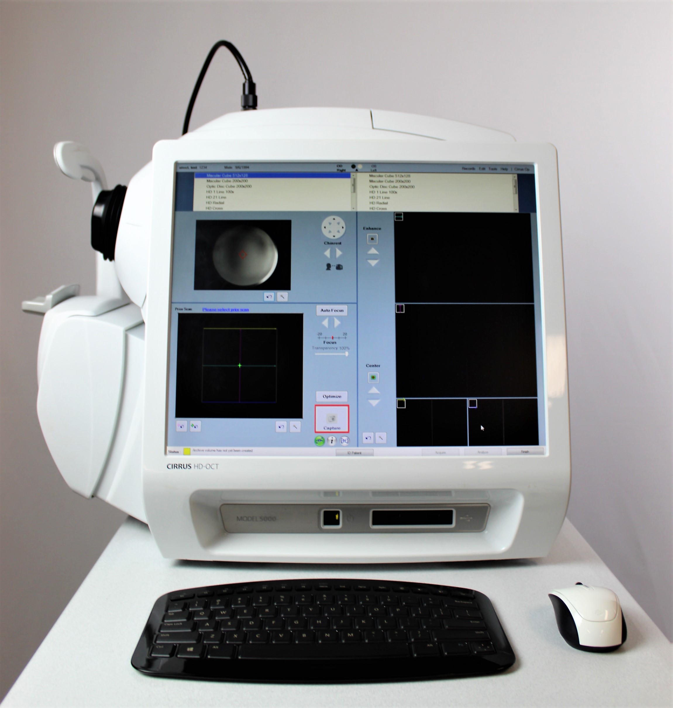

Carl Zeiss Cirrus HD OCT 5000
$ 69.70
Description CARL ZEISS CIRRUS HD OCT 5000 Carl Zeiss Cirrus HD OCT 5000 is configured with the highest resolution visualization capabilities and sophisticated clinical applications such as Advanced RPE analysis to track retinal pigment epithelial integrity and Ganglion Cell Analysis to assess glaucomatous loss in the macula that may not be evident in the peripapillary region. The new FastTrac retinal tracking reduces eye motion artifacts without sacrificing patient throughput with a proprietary scan acquisition strategy, high speed 20 Hz LSO camera, and single-pass alignment scanning. This feature enables the highest resolution B-Scans to be captured in identical locations from visit to visit providing precise assessment of change in targeted pathologies. Features HD Smart Scans FastTrac™ retinal tracking system Macular Thickness OU Analysis Advanced RPE analysis Ganglion Cell Analysis Guided Progression Analysis (GPA) Precision FoveaFinder Macular Thickness and Change Analysis Macular Thickness Normative Data ZEISS AngioPlex OCT Angiography The introduction of ZEISS AngioPlex ushered a new era in retinal care. Now, ultra-clear 3D microvascular visualizations with non-invasive technology can be a routine part of your retina practice. AngioPlex OCT-A allows you to visualize without the use of a contrast agent. Advancing Smart OCT for Anterior Segment, Glaucoma and Retina Anterior Segment Premier Module PanoMap Wide-Field Display Smart HD Scans En face report Great Images & Dense Data Cubes Analyze one pathology from multiple views! Cirrus Enhanced HD Raster Scans are second to none. Capture highly dense data cubes for all the detail you need for analyses and B-scans that help identify even the most subtle pathology. Enhanced High Definition Scans With world-class ZEISS optics and knowledge from over a decade of experience in OCT, Cirrus captures spectacular images second to none. Rather than image averaging, Cirrus uses Selective Pixel Profiling™ to optimize data at each pixel. Judge the results for yourself. Specifications OCT Imaging Methodology: Spectral domain OCT Optical source: Superluminescent diode (SLD), 840 nm Scan speed: 27K- 68K A-scans per second A-scan: 2.0 mm (in tissue), 1024 Axial resolution: 5 μm (in tissue) Transverse resolution: 15 μm (in tissue) Tracking at the speed of CIRRUS FastTrac™ reduces eye motion artifacts without sacrificing patient throughput with a proprietary scan acquisition strategy, high speed 20 Hz LSO camera, and single-pass alignment scanning. With FastTrac, scan at the highest resolution at the same location at each visit. Assessment at the speed of CIRRUS Measurement centering with FoveaFinder and AutoCenter On the macula, the unique FoveaFinder technology ensures the ETDRS and ganglion cell plus inner plexiform layer measurement frameworks are centered on the fovea.